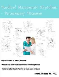
School of Medicine, medical school, Medical School, medical students, medicine, medical student, student, medical education, research, application, Medicine, residency, financial aid, Office, Education, school, the School, Physician, Medical Schools, graduate students
Pulmonary Medicine for Studs
Pulmonary Medicine Notes for Medical Students Studying for the USMLE
Other Studs...
Other Sites
GetResponse
PULMONOLOGY
Obstructive Pulmonary Disease Restrictive Pulmonary Disease
· airway resistance · in lung recoil
· ¯ expiratory flow rate · ¯ in all lung volume
· TLC , ¯ FEV1 / FVC · or N FEV1/ FVC
PFT suggest obstructive pattern ® Diffusing Capacity ¬ PFT suggest restrictive pattern
½
¯ ¯
¯ ¯ ¯ ¯
N/ Near N ¯¯¯¯ N / Near N ¯¯¯
¯ ¯ ¯ ¯
Chronic Bronchitis Emphysema Extrapulmonary Interstitial
Asthma Restriction lung disease
( eg. Kyphoscoliosis, morbid obesity)
- A – a gradient = 150 – 1.25 X PCO2 – PaO2
- Normal ® 5 –15 mmHg
Hypoxemia
N A-a gradient A – a gradient
¯ ¯
Extrapulmonary origin Pulmonary Origin
· PAO2 = % O2 ( 713) – arterial PCO2 / 0.8
· PAO2 = 0.21 ( 713) – 40 / 0.8 = 100 mmHg
· PaO2 = 95 mmHg
· A-a gradient = 5 mmHg
· Atelectasis – most common cause of fever in 1st 24-hrs post operatively
* CXR – best initial diagnostic test for pulmonary diseases
C X R
Calcified Pulmonary Nodule
Low Risk Patient High Risk Patient
(<>50 yrs old, smoker)
C X R every 3 months x 2 yrs Open lung biopsy
Stop follow up, if no growth
C X R
Pleural Effusions
Thoracocentesis (Next Best Step)
¯
LDH & Protein level
Transudate Exudate
LDH effusion <> 200
LDH E / S <> 0.6
Protein E / S < 0.5 < 0.5
- All 3 value must meet to diagnose transudative effusion
- If atleast one criterion is not met, then exudative effusion
placement in patient with parapneumonic effusion
CXR
¯
Atelectasis
¯
Appears on CXR as volume loss / densely consolidated
¯
Bronchoscopy with subsequent removal of mucous plug (Best Treatment / next step)
· Bacterial Pneumonia ® Consolidation ® dull to percussion, vocal framitus
· Pneumothorax ®Hyperresonant to percussion, ¯ Breath sound
· Pleural Effusion ® dull to percussion, ¯ Breath sound, ¯ vocal framitus
· Atelectasis ® dull to percussion, Absent Breath sound, loss of framitus
Pneumothorax ® opposite side
· Deviation of Trachea
Atelectasis (upper lobe) ® same side
· Atelectasis (lower lobe) ® elevation of diaphragm (same side)
· Trachea → Rt & Lt main bronchus → terminal bronchioles → respiratory
bronchioles → alveolar duct (AD) → alveoli
· Foreign body inhalation in child, next step? Direct laryngoscopy and rigid
bronchoscopy to remove foreign body
Asthma : Reversible airway obstruction
˗ Cough induced by forceful expiration (characteristic of airway hyperactivity)
˗ Poor prognosis factors – Pulsus paradoxus, absent breath sounds, decrease
wheezing, cyanosis, bradycardia, normal PaCO2 in acute Asthmatic patient
˗ Best initial therapy – B-agonist(Albuterol)
˗ Exercise induced asthma – beta agonist / chromolyn (mast cell stabilizer)
˗ Acute exacerbation of Asthma – Oxygen (first-step) and Inhaled beta2-agonist
[steroids can be used when beta2-agonist fail. IV steroid is preferred over oral]
[oral steroid is only used if patient is not vomiting]; IV Steroids
˗ PFT should be done for documentation of Asthma
· Asthmatic patient present with c/o worsening of Asthma at night, night cough &
Wheezing, Diagnosis? GERD; next step? proton pump inhibitors (omeprazole,
pentoprazole)
· Subcutaneous emphysema in asthamatic (crepitation over the face & neck), next
step? – CXR [it is benign in asthamatic patient. CXR should be order to rule out
pneumothorax]
· “Silent chest” (absent air entry bilaterally) in Acute asthmatic patient on IV
steroid, next step? Intubation
· Allergic bronchopulmonary Aspergillosis (ABPA) – skin prick test (first step)
should be done in all asthmatic patient who are suspected of having ABPA; Tx:
oral prednisone
· Chronic eosinophilic pneumonia – peripheral infiltrates that are photographic
negative of pulmonary edema on CXR is the pathognomonic feature of chronic
eosinophilic pneumonia – >40% of eosinophils on bronchoalveolar lavage
COPD(Emphysema & chronic Bronchitis) :
· Non-reversible airway obstruction
· ↓ Recoil & ↑ Airway resistance
· Emphysema : cigarette smoking & alpha1-anti-trypsin deficiency (AAT) -
↑ compliance (more dilated alveoli) and ↓ elasticity (failure to keep airway lumen
open – essential for expiration so air trapped during expiration) – Centriacinar
[distended respiratory bronchioles – air trapped in AD & alveoli] Panacinar
[distended whole respiratory unit (Resp bronchioles, AD & alveoli) – air trapped
in whole unit]
· Chronic Bronchitis : productive cough for at least 3 months for 2 consecutive
years – smoking cigarette & cystic fibrosis - ↑mucus in bronchi obstruct terminal
bronchioles & narrowing of lumen due to chronic inflammation and fibrosis
Emphysema Hyperinflation of bilateral lung fields
With flattening of diaphragm, small
CXR – Size heart
Chronic Bronchitis Increased pulmonary markings
· PFT – best diagnostic test
· FEV1 – Single most important factor to determine prognosis of COPD
· Treatment – Smoking cessation (Best treatment to slow progression)
· Acute Exacerbation → Oxygen, Systemic steroid, Antibiotics (Should cover
H.influenzae & Pneumococcus), Bronchodilators
· Out patient Tx option → Ipratropium (1st line drug) & Home O2
Bronchodilators (2nd line drug)
Vaccination [Influenza (yearly)]
· First step in management of massive hemoptysis Rigid bronchoscopy (not flexible)
· Copious in amount & foul smelling sputum, initial test? CRX; next step?
CT chest [Causes: Bronchiectasis, Lung Abscess, Anaerobic Pneumonia]
■ Lung Abscess : Alcoholic, Extremely bad odor (like decomposing dead animal)
TB : Alveolar macrophage → CD4+ T-cells → Macrophage release IL-12
(stimulates TH1 cells) and IL-1 (fever; activate TH1 cells) → TH1 release IL-2 (self
stimulation of TH1) and ᵧ interferon (activate macrophage to kill tubercular bacilli) → Inflammatory mediators release from macrophage are responsible for
tissue damage (no endotoxin or exotoxin) → Lipid from tubercular bacilli leads to
caseous necrosis
Bronchiactasis:
- Permanent dilation of bronchi & bronchioles
- Chronic infection [gram(-) organisms] [destruction of cartilage & elastic tissues]
- Persistent cough with purulent copious sputum production, wheezes, crackles
- Chest CT – Best noninvasive test
- Treatment → Bronchodilators, chest physical therapy, postural drainage, and
Antibiotics
- Vaccination (Pneumococcal – every 5 yrs & Influenza – yearly)
Idiopathic pulmonary Fibrosis :
- Involve only lung except clubbing
- Unknown etiology, occur in 5th decade
- CXR – Reticular / Reticulonodular disease
- Chest CT – ground glass appearance
- PFT- restrictive pattern
- Treatment – steroid with / without Azathioprine (help in only 20% of pt)
Sarcoidosis :
- 20 – 40 yrs. Old women
- Presence of nonspecific non-caseating granuloma in the lung and other organs
- CXR – bilateral hilar adenopathy (90% of cases)
- Hypercalcemia (↑ 1-α-hydroxylase by macrophage leads to ↑ Vit–D)
[Hypercalciuria is occur in around 50% of cases whereas Hypercalcemia is seen
in around 10-20% of cases of Sarcoidosis]
- ↑ ACE (60 % of patients)
- Decrease cellular immunity [low helper/suppressor T-cell ratio] and activation of
humoral immunity [increase CD4/CD8 ratio]
- Ophthalmoscopic examination (uveitis & conjunctivitis - >25% of the cases)
- Treatment – systemic steroids when it involve uveitis / CNS / Hypercalcemia
- Tx of Hypercalcemia in Sarcoidosis – hydration + glucocorticoids
- Pulmonary pathology in Sarcoidosis inflammatory granuloma
- Pulmonary pathology in Systemic Sclerosis (Scleroderma) interstitial fibrosis
Pneumoconiosis :
- CXR – small irregular opacities, interstitial densities, ground glass appearance,
honey combing
- Asbestosis → H/O exposure, usually involve lower lung fields
- CXR – diffuse /local pleural thickening, pleural plaques, calcification at the level
of diaphragm
- Lung biopsy – barbell shaped asbestos fiber (Best diagnostic test)
- ↑↑↑ Risk of Bronchogenic CA
- ↑ Risk of pleural / peritoneal mesothelioma
Coal miner’s / coal worker’s pneumoconiosis (CWP) :
- Usually involve upper half of lung
- Increase Levels of IgA, IgG, C3 , anti-nuclear Ab, RF
- Caplan syndrome – Rheumatoid nodule in the periphery of the lung in a patient
with RA & CWP
Silicosis Hyaline nodule, usually involve upper lobe
- Strong association with TB , Pt should go yearly PD & if PPD >10mm then INH
for 9 months
Pulmonary Thromboembolism :
- Sudden onset of dyspnea along with tachycardia
- ECG Right Axis Deviation
- h/o long term immobility
- Ventilation – perfusion (V /Q) scan (Best initial test)
- Pulmonary Angiogram (most accurate test)
- V/Q scan → Doppler U/S of lower limb or CT angiogram of chest → Pulmonary angiography
- Treatment : continuous Heparin therapy x 5 days + Warfarin x 6 months. If Hemodynamically unstable → THROMBOLYTICS, EMBOLECTOMY (IF THROMBOLYTICS ARE CONTRAINDICATED )
- Pregnant patients → low molecular weight Heparin x 6 months
· Pleuritic chest pain (pain increase on inspiration), tachycardia & dyspnea in patient on contraceptive pills, diagnosis?
Pulmonary embolism / infarction
Adult Respiratory Distress Syndrome (ARDS) :
- ↑ Permeability of the alveolar – capillary membrane & Pul. edema
- Alveolar macrophage → cytokines → Neutrophil → damage capillary membrane
- CXR – diffuse interstitial infiltrates; whiteout of both lung fields
- Swan-Ganz Catheter – normal cardiac output & capillary wedge pressure
↑ Pulmonary artery pressure
˗ Mx of ARDS: PEEP (around 8-9 cmH2O), High oxygen and low tidal volume; Oxygen flow should be decrease after patient improve to prevent oxygen toxicity
Sleep Apnea : Daytime Somnolence
- Obstructive sleep Apnea → floppy airway, obese patient
- Central sleep Apnea → inadequate ventilatory drive
- Treatment → Acetazolamide, progesterone and supplemental O2
Bronchogenic Carcinoma :
- Squamous cell CA → Centrally located → Hypercalcemia – PTH-like substance
- Small cell CA → Centrally located → SIADH, Eaton-Lambert, Venocaval obstruction Syndrome, Horner’s syndrome
- Large Cell CA → Peripherally located
- Adenocarcinoma → peripherally located → Pleural effusion with high hyaluronidase level in effusion fluid. Bronchoalveolar CA is subtype
- Popcorn calcification, concentric, central, or diffuse homogenous calcification on CXR is suggestive of a benign pathology of pulmonary nodules
· Diagnosis : Sputum Cytology (SCC - >80%), Bronchoscopy (>90% for centrally located)
- Needle Aspiration Biopsy → peripheral nodule with effusion
- Treatment : Resection (when possible), etoposide & platinum for small CA,
- Non-small cell CA – Cyclophosphamide, Adriamycin and platinum
- Effusions can be sclerosed with Tetracycline
· Pancoast tumor – Horner’s syndrome, Phrenic N involvement [Chest movement asymmetry] [a dangerous sign in patient with Pancoast tumor]; Radiation therapy is the treatment of choice for Pancoast tumor with distant metastasis
· Horner’s syndrome in smoker, next step? – CXR (to rule out lung CA)
Studly Overview
Sinusitis
Allergic Rhinitis
Infectious Parotitis
Aphthous Stomatitis
Hand-Foot-Mouth Disease
Influenza
Sleep Apnea Syndrome
Streptococcal Sore Throat
Scarlet Fever
Diphtheria
Acute infectious Laryngitis
Acute Laryngotracheiitis
Congenital Tracheomalacia
Mediastinal Diseases
Acute Mediastinitis
Mediastinal Masses
Sarcoidosis
Squamous Cell Carcinoma
Bronchiestasis
Bronchopulmonary Dysplasia
Cystic Fibrosis
Chronic Obstructive Pulmonary Disease
Asthma
Bronchiestasis
Cystic Fibrosis
Bronchiolitis
Bronchiolitis Obliterans with Organizing Pneumonia
Alveolar Proteinosis
Alveolar Cell Carcinoma
VASCULAR
Goodpasture's Syndrome
Wegener's Granulomatosis
Pulmonary Arteriovenous Fistula
Pulmonary Embolism
Treatment: IV heparin followed by coumadin for 6 months
Pulmonary Hypertension
Cor Pulmonale
Pneumonia
Tuberculosis
Lung Abscess
Diffuse Interstitial Lung Disease
Desquamative Interstitial Pneumonitis
Pulmonary Hemosiderosis
Occupational Lung Diseases
Emphysema
Large Cell Carcinoma
DISEASES OF THE PLEURA
Pneumothorax
Types
- Primary Spontaneous Pneumothorax
Chylothorax
Asbestosis/Mesothelioma
Chest Wall Disorders
Kyphoscoliosis
Sleep Apnea Syndrome
Treatment: CPAP, surgery
Sarcoidosis
Characterized by noncaseating granulomas
Treatment: steroids
Bronchiolitis
RSV accounts for most cases
O2 treatment
Pleural Effusion
Typical: Streptococcus pneumoniae
Atypical
